Spine Imaging
&
Disease Prediction

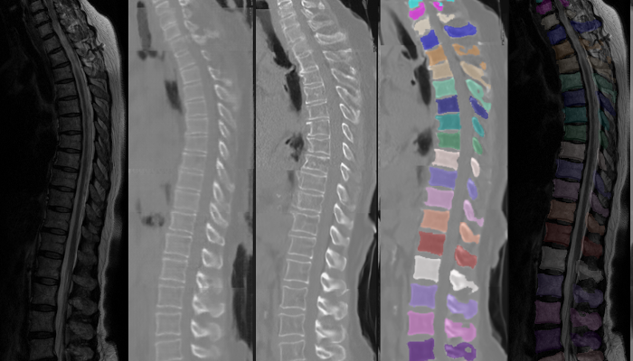
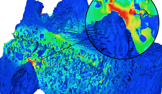
We study new imaging techniques in CT and MRI for quantitative imaging of the spine. We develop automated evaluation algorithms including artificial intelligence, deep learning and biomechanic modeling to predict disease progression and get new pathophysiologic insights. Currently we focus on osteoporosis, back pain, degenerative spine diseases and inflammatory disorders like multiple sclerosis. We work with in-house datasets as well as with data from GNC and SHIP studies

Projects


Image analysis in epidemiologic studies

We develop AI-based methods to automatically analyze and quantify MR images of the spine. We currently work on methods to translate MRI to CT to use existing labeling and segmentation methods. Our goal is to analyze large datasets from GNC to better understand sources of back pain.

Biomechanic modeling

To better understand back pain and surgical outcome, we combine quantitative parameters from different imaging modalities (X-rays, MRI, CT) in one biomechanical model, based on multibody simulations (MBS) on a large scale and finite element models (FEM/FCM) on a small scale.

Spinal Multiple Sclerosis

We investigate different techniques to automatically assess spinal lesion load and its clinical relevance for the individual patient.

We closely collaborate with the group of Prof. Mühlau and Prof. Cohen-Adad to further increase functionaliy and robustness of the spinal cord toolbox.

ANDUIN

A fully automated bone segmentation software

Anduin is a freely available web-based application able to fully automatically segment the spine in clinical CT scans. Our spin-off "Bonescreen"  has further developed the segmentation code (contact them directly for scientific collaborations) and is dedicated to establish a fully automated opportunistic osteoporosis diagnosis in clinical routine.
Try out Bonescreen!

The VerSe Challenges

Driving forward an interest for fully automated segmentation of the spine in the research community, we publicly released voxel-level-annotated CT data of more than 300 patients and organised the VerSe (Large Scale Vertebrae Segmentation) challenge series MICCAI 2019 and 2020. See the resulting MedIA paper (or ArXiv here)

Opportunistic Screening in Osteoporosis

Together with Bonescreen, we validate and develop automated techniques for opportunistic BMD measurements in any CT dataset. We also implemented automatic fracture detection and prediction algorithms using artificial intelligence. Currently, we focus on the spine.

Selected Publications


Somatosensory evoked potentials and magnetic resonance imaging of the central nervous system in early multiple sclerosis. Wuschek A, J Neurol. 2022 Oct 7. 

Validation of a Patient-Specific Musculoskeletal Model for Lumbar Load Estimation Generated by an Automated Pipeline From Whole Body CT. Lerchl T, Front Bioeng Biotechnol. 2022 Jul 11;10:862804.

Beyond mean value analysis - a voxel-based analysis of the quantitative MR biomarker water T2 in the presence of fatty infiltration in skeletal muscle tissue of patients with neuromuscular diseases. Schlaeger S, NMR Biomed. 2022 Dec;35(12):e4805.

Patient-Specific Finite Element Modeling of the Whole Lumbar Spine Using Clinical Routine Multi-Detector Computed Tomography (MDCT) Data-A Pilot Study. Rayudu NM, Biomedicines. 2022 Jun 30;10(7):1567.

[Postoperative imaging of the musculoskeletal system - spine]. Schlaeger S, Kirschke JS. Radiologie (Heidelb). 2022 Oct;62(10):851-861.

Level-Specific Volumetric BMD Threshold Values for the Prediction of Incident Vertebral Fractures Using Opportunistic QCT: A Case-Control Study. Dieckmeyer M, Front Endocrinol (Lausanne). 2022 May 20;13:882163.

Automated Opportunistic Osteoporosis Screening in Routine Computed Tomography of the Spine: Comparison With Dedicated Quantitative CT. Sollmann N, J Bone Miner Res. 2022 Jul;37(7):1287-1296.

Multiple sclerosis lesions and atrophy in the spinal cord: Distribution across vertebral levels and correlation with disability. Bussas M, Neuroimage Clin. 2022;34:103006.

Proposed diagnostic volumetric bone mineral density thresholds for osteoporosis and osteopenia at the cervicothoracic spine in correlation to the lumbar spine. Rühling S, Eur Radiol. 2022 Sep;32(9):6207-6214.

Anatomy-Aware Inference of the 3D Standing Spine Posture from 2D Radiographs. Bayat A, Tomography. 2022 Feb 11;8(1):479-496.

A computed tomography vertebral segmentation dataset with anatomical variations and multi-vendor scanner data. Liebl H, Sci Data. 2021 Oct 28;8(1):284.

Automated detection of the contrast phase in MDCT by an artificial neural network improves the accuracy of opportunistic bone mineral density measurements. Rühling S, Eur Radiol. 2022 Mar;32(3):1465-1474.

Epidemiology and reporting of osteoporotic vertebral fractures in patients with long-term hospital records based on routine clinical CT imaging. Löffler MT, Kallweit M, Niederreiter E, Baum T, Makowski MR, Zimmer C, Kirschke JS.Osteoporos Int. 2022 Mar;33(3):685-694.

MR-based proton density fat fraction (PDFF) of the vertebral bone marrow differentiates between patients with and without osteoporotic vertebral fractures. Gassert FT, et al. Osteoporos Int. 2022 Feb;33(2):487-496.

VerSe: A Vertebrae labelling and segmentation benchmark for multi-detector CT images. Sekuboyina A, et al. Med Image Anal. 2021 Oct;73:102166. 

Prediction of incident vertebral fractures in routine MDCT: Comparison of global texture features, 3D finite element parameters and volumetric BMD. Dieckmeyer M, Eur J Radiol. 2021 Aug;141:109827.

A Vertebral Segmentation Dataset with Fracture Grading. Löffler MT, Radiol Artif Intell. 2020 Jul 29;2(4):e190138.

Automatic opportunistic osteoporosis screening in routine CT: improved prediction of patients with prevalent vertebral fractures compared to DXA. Löffler MT, Eur Radiol. 2021 Aug;31(8):6069-6077.

Opportunistic Osteoporosis Screening Reveals Low Bone Density in Patients With Screw Loosening After Lumbar Semi-Rigid Instrumentation: A Case-Control Study. Löffler MT, Front Endocrinol (Lausanne). 2021 Jan 11;11:552719.

Group


...
Thomas Baum
Co-Principal Investigator
...
Anjany Sekuboyina
PhD Student
...
Tanja Lerchl
PhD Student
...
Robert Graf
PhD Student
...
Hendrik Möller
PhD Student
...
Malek Husseini
PhD Student
...
Kati Nispel
PhD Student
...
Sebastian Rühling
MD
...
Egon Burian
MD
...
Sarah Schlaeger
MD
...
Nico Sollmann
MD
...
David Schinz
MD
...
Maximilian T. Löffler
MD
...
Daria Bischl
Cand. med.
...
Jonas Dittmann
Cand. med.
...
Mareike Hebell
Cand. med.
...
Charlotte Kampf
Cand. med.
...
Benjamin Keinert
Cand. med.
...
Magdalena Griesbauer
Cand. med.
...
Matthias Bruckbauer
Cand. med.
...
Stefan Ruschke
Cand. med., Dr. rer. nat.
...
Philipp Sperl
MD
...
Viktoria Thierauf
Cand. med.
...
Rikarda Wurm
Cand. med.
...
Theresa Zarth
Cand. med.
...
Sofiia Pazynych
Cand. Med.
...
Magaly Gutbrod-Fernandez
Cand. Med.
...
Isabel Seeger
Cand. Med.
...
Eva Niederreiter
Cand. Med.

Alumni


...
Michael Dieckmeyer
MD
...
Amir Bayat
PhD Student
...
Mohamed Elhaddad
PhD Student
...
Alina Jakob
MD
...
Anna-Lena Grau
MD
List of completed thesis of (previous) group members

Collaborators


Funding


...
The European Research Council (ERC) under the European Union’s Horizon 2020 research and innovation programme
(iBack —ERC-2014-STG) 2015-2020
(Bonescreen - 2020 PoC) 2020-2021
(iBack-epic — ERC-2021-CoG) 2022-2027
...
The German Research Foundation 2010-2024
...
The German Federal Ministry for Economic Affairs and Energy 2012-2015 and 2021-2023


jan.kirschke@tum.de   

Abteilung für Neuroradiologie Klinikum rechts der Isar

Technische Universität München, Ismaninger Str. 22, 81675 München

V E R S E

The Large Scale Vertebrae Segmentation Challenge Series


VerSe`19

Key highlights

  • Held at MICCAI 2019, Shenzhen, China.
  • Public release of an unprecedented 160 spine CT scans with voxel-level vertebral annotations.
  • 500+ data download requests!
  • Benchmarked 20 labelling and segmentation algorithms.
  • Dataset released under CC BY-SA 2.0 license.

VerSe`20

Key highlights

  • Held at MICCAI 2020, Lima, Peru.
  • Public release of an unprecedented 300 spine CT scans with voxel-level vertebral annotations.
  • Multi-center, multi-scanner dataset.
  • Focus on peculiar spinal anatomies incl. transitional vertebrae.
  • Dataset released under CC BY-SA 4.0 license.